Overview
Electrical Impedance Tomography (EIT) serves as a non-invasive imaging method with significant potential in scientific and clinical domains, particularly for bedside monitoring of lung ventilation, perfusion, and regional lung function. EIT examinations involve strategically placing electrodes around the chest circumference to capture relevant phenomena. Typically integrated into belts or strips, these electrodes align in a transverse plane. Small alternating electrical currents pass through electrode pairs during the examination, measuring resulting voltages across the remaining electrodes.
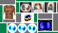
Employing adjacent electrode pairs for current application and voltage measurements, mathematical reconstruction methods then generate tomographic lung images based on these measured voltages, illustrating the patient's current ventilation status.
Current research at ITeM
- Integrating data from other devices (e.g., x-ray computed imaging, motion tracking systems, spirometry) into EIT image reconstruction.
- Developing novel, rapid image reconstruction algorithms suitable for 3D-EIT image generation.
- Exploring the nuances of EIT usage in patients with obstructive lung diseases.
- Detailed investigation of the screw insertion and tightening processes using FEM analysis
- Developing algorithms to streamline EIT data interpretation in clinical contexts, such as with ARDS or COVID-19 patients.
- Ludwig-Maximilians-Universität Munich
- University of Canterbury
- Maastricht University Medical Center+
- Budapest University of Technology and Economics